Figure 3.
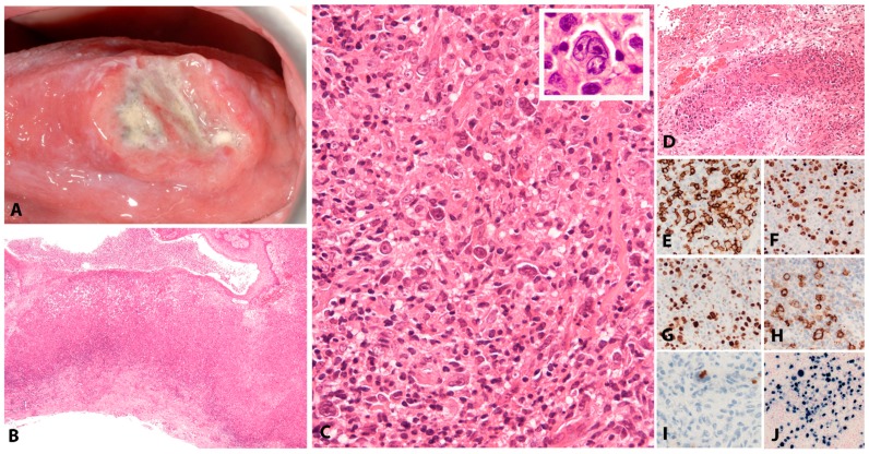
EBV positive mucocutaneous ulcer. (A) EBV MCU on the tongue—a shallow ulcer with raised edges and necrotic debris in the centre. The clinical suspicion is often one of squamous cell carcinoma. (B) The ulcer is well circumscribed and at the base shows a rim of darker staining small lymphocytes (H&E, 10×). (C) On higher magnification, it comprises a polymorphous mixture of lymphoid cells of variable sizes, many with HRS-cell features (H&E, 200×) (inset) in a lymphohistiocytic background (H&E, 600×). (D) Angioinvasion is frequently seen. (E) The lesional cells are significantly positive for CD20, which highlights variable cell size. (F) There is strong expression of PAX5. (G) OCT2 is also strongly positive. (H) Most cells express CD30. (I) There is frequent co-expression of CD15 (immunohistochemistry, D, 100×; E, F, G, H, 200×; I, 400×). (J) EBER is abundantly positive, highlighting variability in nuclear sizes of the lesional cells (in-situ hybridization, 200×).

