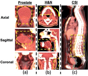
Figure 1.

The virtual deformed images for the three clinical cases. Each of the images represents only one slice of axial, sagittal, and coronal view of a 3D CT data set. For CSI case, only the sagittal view is shown. The original and simulated deformation images are fused together to show the difference in deformation (highlighted in the yellow box): (a) prostate case with bladder filling, (b) head and neck case with significant weight loss, deformation includes head twisted by a small angle, relative smaller oral cavity closure even with a bite‐block, and noticeable narrower neck region; (c) cranial spinal irradiation case at prone position first then with lower spine IMRT boost at supine position with less curved spine.
