Figure 1.
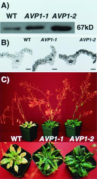
AVP1 overexpression increases salt tolerance. (A) Western blot of membrane fractions from wild-type and transgenic plants. Protein (10 μg) from total membrane fractions isolated from shoots of four wild-type and four plants of each of the transgenic lines (AVP1-1 and AVP1-2) was separated by 10% SDS/PAGE. Four SDS/PAGE gels were transferred and immunoblotted with antibodies raised against a keyhole limpet hemocyanin-conjugated AVP1 peptide (see Materials and Methods). AVP1 protein was detected by chemiluminescence. The photograph corresponds to one of four immunoblots. WT, wild type. (B) Immunocytochemical localization of AVP1 protein in wild-type, AVP1-1, and AVP1-2 transgenic plants. Sections of rosette leaves were probed with the AVP1 antibody. Antibody binding was detected with NBT/BCIP as substrate (see Materials and Methods). (Bars, 200 μm.) (C) Salt treatment of wild type and two AVP1-overexpressing transgenic lines. Plants were grown on soil in a 16-h light/8-h dark cycle and treated as described in Materials and Methods. The photograph shows plants at the 10th day after treatment with 250 mM NaCl (see Materials and Methods).

