FIG 2.
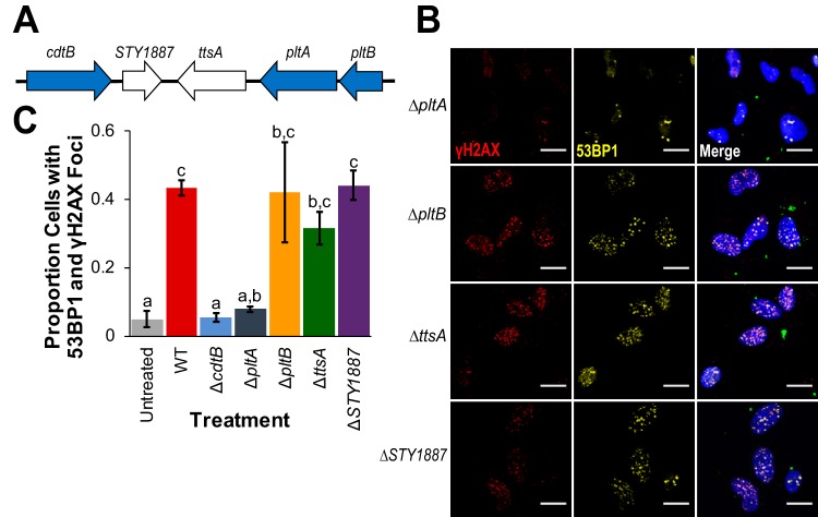
pltB, ttsA, and STY1887 are not required for S-CDT-mediated DNA damage in HIEC-6 cells. HIEC-6 cells were infected with S. Javiana strains harboring single-gene deletions in the S-CDT islet. Immunofluorescence staining was performed for DNA damage response foci γH2AX and 53BP1. (A) Organization of the S-CDT islet in S. Javiana. Genes shown in blue represent genes encoding S-CDT subunits; genes shown in white are contained within the islet but do not encode protein products that constitute part of the S-CDT holotoxin. (B) Representative images of cells infected with wild-type S. Javiana and S-CDT single gene deletion strains (both colored green in the merged image); HIEC-6 cell nuclei are shown in blue. (C) Quantification of the proportions of cells with at least four 53BP1 foci that colocalized with γH2AX foci. Treatments that do not share letters have significantly different (P < 0.05) proportions of cells with 53BP1 foci that colocalized with γH2AX foci. Data represent three independent experiments. P values were corrected, to account for multiple testing, using Tukey’s HSD test. Error bars represent standard errors of the means.

