
FIG 3 .
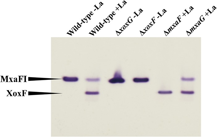
MDH activities in mutants generated in this study compared to wild-type strain activities. MDH enzymes were visualized through activity staining of the gel, after separation by electrophoresis in a gradient (4 to 25%) polyacrylamide gel. La was supplied at 30 μM where indicated. Cell extracts were adjusted to approximately 3 mg/ml protein, and 10 μl of extract was applied in each lane of the gel.