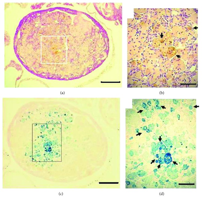
Figure 1.
Hemosiderin in the aged C56BL/6 ovary. HE stain (a, b) and Perls stain (c, d) of a virgin ovary 20.6 months old. Arrows in (b) indicate brown-yellowish granules of hemosiderin. Arrows in (d) show hemosiderin laden macrophages (HLMs). Bars in (a) and (c) = 200 μm; bars in (b) and (d) = 20 μm.

