
Figure 5. RyR1 moving parts.
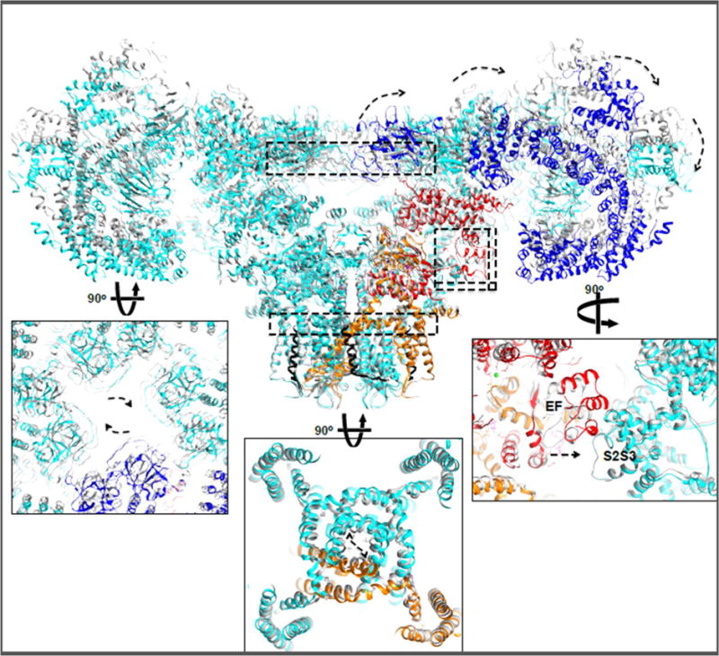
Upon Ca2+ and ATP binding dilated channel pore aperture can be observed coupled to exo-rotation of the cytoplasmic shell and clockwise translation of the NTD cytoplasmic vestibule. Dashed arrows represent the apparent direction of movements from closed to open states. Similar conformational changes were observed in in the presence of PCB95 or ruthenium red. Curiously, an ~8 Å translation of the EF-hand pair towards the S2–S3 helical bundle is observed also in the RyR1 ‘primed’ state. Insets show enlarged views of the structures at the indicated dashed rectangles.