Figure 4.
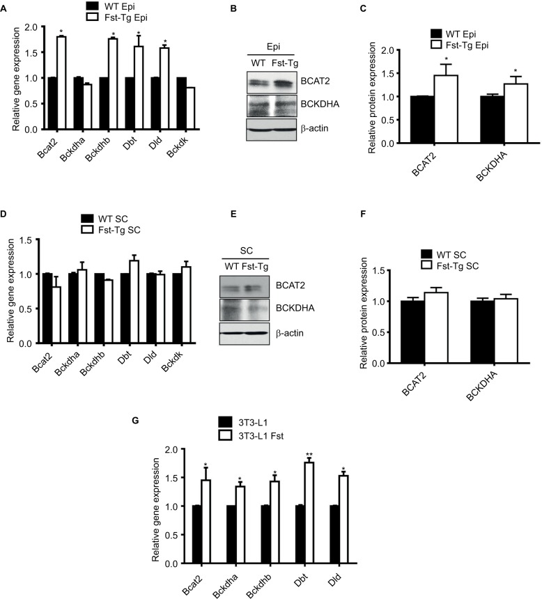
Analysis of key genes and proteins involved in BCAA catabolic pathways. (A) Quantitative gene expression analysis of mitochondrial Bcat2 and key BCKDH complex enzymes in Epi adipose tissues isolated from male 10-week-old WT and Fst-Tg mice. Western blot analysis using 100 µg total cell lysates (B) and densitometric quantitation (C) of BCAT2 and BCKDHA proteins in Epi adipose tissues isolated from male WT and Fst-Tg mice. (D) Real-time quantitative gene expression analysis of mitochondrial Bcat2 and key BCKDH complex enzymes in SC adipose tissues isolated from male WT and Fst-Tg mice. Western blot analysis (E) and densitometric quantitation (F) of BCAT2 and BCKDHA proteins in SC adipose tissues isolated from male WT and Fst-Tg mice. Data are expressed as mean ± SD. *P≤0.05 (n=3). (G) Real-time quantitative gene expression analysis of mitochondrial Bcat2 and key BCKDH complex enzymes in differentiating 3T3-L1 and 3T3-L1 Fst cells. *P≤0.05, **P≤0.01 (n=3).
Abbreviations: WT, wild type; Fst-Tg, follistatin transgenic; Epi, epididymal; BCAT, branched chain aminotransferase; BCKDHA, branched chain ketoacid dehydrogenase; BCAA, branched chain amino acid; SC, subcutaneous.

