
Figure 1.
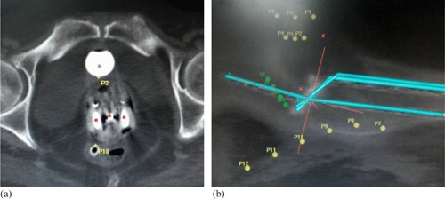
CT Scan images showing the marking of bladder and rectal points: (a) axial CT image showing ICBT applicator with central tandem and two ovoids where the bladder point can be seen on the posterior surface of Foley's bulb and the rectal point can be seen on the anterior surface of marker tube inserted into the rectum; (b) sagittal CT image showing the reconstructed ICBT applicator, multiple bladder points anteriorly and rectal points posteriorly.