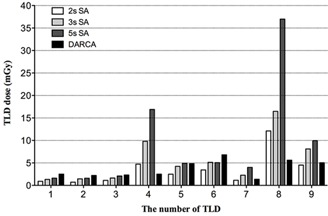
Figure 3.

Skin dose distribution in the thermoluminescent dosimeter arrays separated by different angiographic protocol. 2 s, 3 s, and 5 s cine time was applied in each projection during the 2 s, 3 s, and 5 s SA, which represents evaluating normal vessels, uncomplicated, and complicated coronary lesions in clinical practice, respectively. TLD = thermoluminescent dosimeter; SA = standard coronary angiography; DARCA = dual‐axis rotational coronary angiography.
