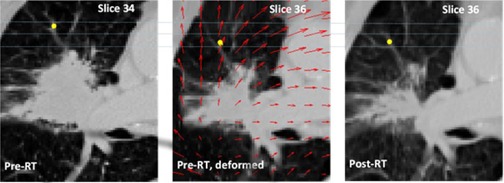
Figure 1.

Representative image for a patient in Group 1 showing a landmark in the pre‐RT scan (left image) and post‐RT CT scan (right image, after rigid registration), the deformed pre‐RT CT scan is shown in the middle. Notice that there is also a difference in craniocaudal distance indicated by the slice numbers. Originally the landmarks in pre‐ and post‐RT scan were two slices apart, but after deformation they are located on the same slice. The deformation field is indicated by red arrows in the deformed scan. Note that the arrows point in the opposite direction of the displacement vectors. The landmark distance was reduced from 14 mm to 3 mm after deformable registration.
