Figure 3.

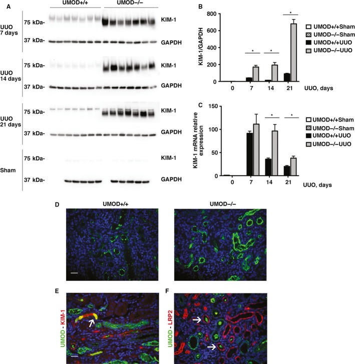
The tubular injury marker KIM‐1 is higher in mice lacking in UMOD during obstructive nephropathy. By western blotting, kidney KIM‐1 protein levels were increased after UUO (n = 5 for sham groups; n = 7 for UUO groups) at all time‐points (days 7, 14, and 21); the levels were significantly higher in the UMOD−/− groups. Results are corrected for protein loading using GAPDH levels. (A, B). Kidney KIM‐1 mRNA levels measured by real‐time PCR (n = 3 for sham groups; n = 5 for UUO groups) were significantly higher on UUO days 14 and 21 (C). Representative day 14 immunofluorescence photomicrographs illustrate more intense KIM‐1 staining in the UMOD−/− kidney (D). KIM‐1 staining was concentrated along the apical membrane with lesser amounts in tubular lumina; the latter more evident in the UMOD+/+ mice. Brightly stained ribbon‐like KIM‐1 + bands, frequently observed along the apical membrane in the UMOD‐/‐ mice, were much less common in the UMOD+/+ mice. Dual staining for UMOD (green) and KIM‐1 (red) in a UMOD+/+ day 14 kidney illustrates an area of tubular intraluminal staining for both proteins (yellow; highlighted by the arrow) (E). Dual staining for UMOD (green) and the proximal tubular apical membrane protein LRP2 (red) in a UMOD+/+ day 14 kidney illustrates the presence of luminal UMOD deposits surrounded by LRP2 + tubules (arrows) (F). Bar graphs represent means +1SEM; * indicates a P value < 0.05. Photomicrograph magnification: ×40. Scale bars: 25 μm.
