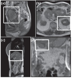
Figure 1.

Sample motion images containing (a) a sagittal view of bladder (S1), (b) an axial view of kidney (S2), (c) a sagittal view of liver tumor (S3), and(d) a coronal view of duodenum (S2). The white box indicates the reduced field of view (FOV) used in the segmentation.
