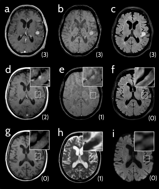
Fig 2. Analysis of pre-diagnosis cMRIs.
69-year-old woman with a melanoma brain metastasis (MBM) that was detectable in an examination prior to the one that led to diagnosis. Conspicuity scores are indicated in brackets below the lesions. (a-c) Axial cMRI scans of a patient with newly diagnosed MBM. (a) Contrast-enhanced T1-weighted image (ceT1w): conspicuity score (CS) 3. (b) Susceptibility-weighted image (SWI), CS 3. (c) Fluid-attenuated inversion recovery (FLAIR) image: CS 3. (d-i) Axial cMRI scans of the pre-examination (ca. 9 weeks earlier).(d) CeT1w: CS 2. (e) SWI: CS 1. (f) FLAIR: CS 0. (g) Non-enhanced T1-weighted image: CS 0. (h) T2-weighted image: CS 1. (i) Diffusion-weighted image (TRACE): CS 0.

