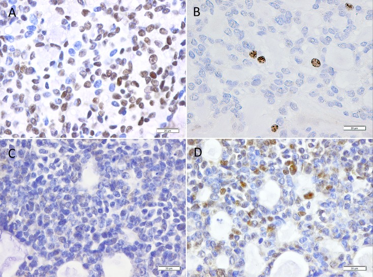
Fig 2. Representative microscopic images of tumor samples from 2 patients with an ACC.
A) shows a tumor sample with a high SOX2 expression using immunohistochemistry (in 4x and 40x magnification) and B) an almost absent expression of ki67. C) shows a tumor sample with no detectable SOX2 expression and D) with elevated levels of ki67.

