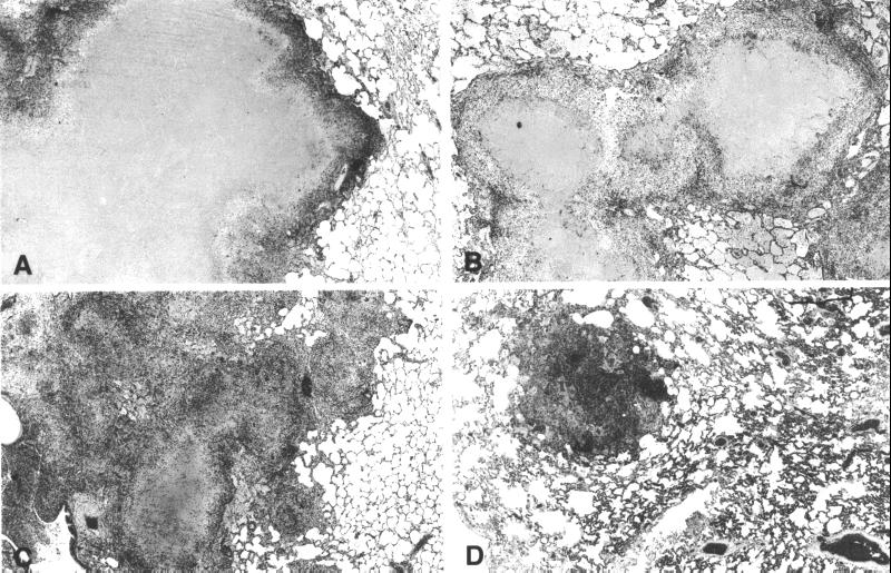
Figure 3.
Histopathology of lungs at week 9 of TB infection in nonvaccinated (A) and bacillus Calmette–Guérin-vaccinated (B) rhesus and nonvaccinated (C) cynomolgus monkeys showing large granulomas with caseous necrosis (A, B, and C). In vaccinated cynomolgus monkeys (D), only a few largely fibrotic granulomas are evident (hematoxylin/eosin staining, original magnification ×6.6).

