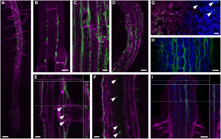
Fig 4. Colonization of Arabidopsis seedlings with GFP-expressing SA187 visualized by confocal microscopy.
(A) Root colonization of agar-grown seedlings starts in the elongation zone. Large colonies then occur in the differentiation zone. MIP; bar = 100 μm. (B) Colonies first established themselves in grooves between root epidermal cells. MIP; bar = 10 μm. (C) Large colonies in the differentiation zone grow out from the grooves. MIP; bar = 10 μm. (D) Root colonization of soil-grown seedlings exhibit a more random pattern in comparison to agar-grown seedlings. MIP; bar = 50 μm. (E) Lateral root emergence allows SA187 to enter the root and colonize the lateral root base (marked by arrowheads). A selected confocal section from a Z-stack with top and side orthogonal views. Bar = 20 μm. (F) Scattered SA187 colonies occur inside the root tissues in two-week-old seedlings (marked by arrowheads). A single confocal section. Bar = 20 μm. (G) In cotyledons, SA187 colonizes grooves between epidermal cells (left side) as well as the extracellular space between mesophyll cells (right side; marked by arrowheads). A single oblique confocal section is shown. Bar = 20 μm. (H) SA187 colonization of the hypocotyl epidermis. MIP; bar = 20 μm. (I) SA187 cells enter hypocotyl via stomata, move freely among hypocotyl cells and occasionally establish colonies inside. A selected confocal section from a Z-stack with top and side orthogonal views. Bar = 50 μm. Green–SA187-GFP; Magenta–cells walls (propidium iodide labeling); Blue–chloroplasts (autofluorescence); MIP–maximum intensity projection of a confocal Z-stack.

