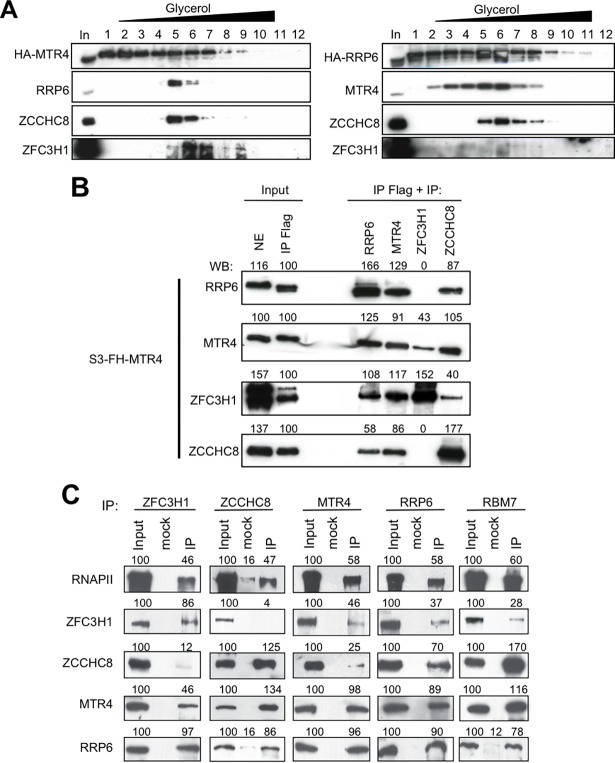
Fig 2. Identification of RRP6- and MTR4- containing complexes.
(A) MTR4- or RRP6-containing complexes were separated by gradient sedimentation analysis followed by immunoblotting using the indicated antibodies. (B) Extracts of cells stably expressing Flag-HA-MTR4 were first immunoprecipitated using anti-Flag then re-immunoprecipitated using the antibodies indicated on the figure. Nuclear extract (NE), anti-Flag immunoprecipitate and the Re-IPs were analyzed by Western blotting using the antibodies indicated. Band intensities quantified by ImageJ are shown above the blots. Samples of the first IP (input IP Flag) were set to 100%. (C) Co-immunoprecipitation analysis of ZFC3H1, ZCCHC8, MTR4, RRP6 and RBM7 was performed using the antibodies indicated on the figure. Values shown above the blots represent the intensity of the bands relative to input samples, which were set to 100%. See also S2 Fig and S3 Fig.

