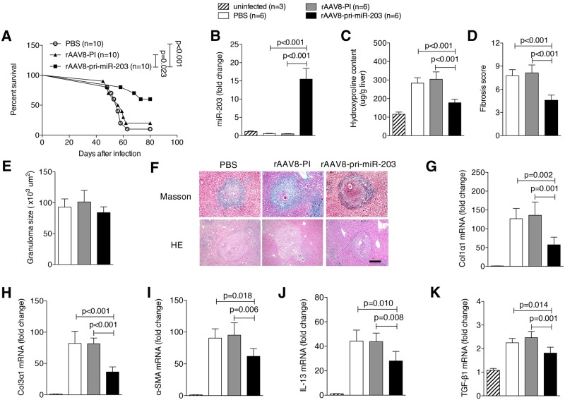
Fig 1. Elevation of miR-203-3p protects hosts from lethal schistosome infection by relieving type 2 pathology.
(A) Mice were infected percutaneously with 30 S. japonicum cercariae at day 0 and treated with rAAV8-PI (n = 10) or rAAV8-pri-miR-203-3p vectors (n = 10) at a dose of 1×10¹¹ virus genomes, or PBS (n = 10) by tail vein injection at day 10 post-infection. The animals were subjected to an 80-day survival study. Data are representative for three independent experiments. Survival between infected groups was compared by Kaplan–Meier survival curves with log-rank test. (B-K) Mice were infected percutaneously with 16 S. japonicum cercariae at day 0 or remained uninfected. Infected mice received rAAV8-PI or rAAV8-pri-miR-203-3p vectors at a dose of 1×10¹¹ virus genomes or PBS by tail vein injection at day 10 post-infection. Liver samples were collected at day 42 post-infection. qPCR analysis of levels of miR-203-3p in the liver samples (B). Striped bars, uninfected mice (n = 3); white bars, mice receiving PBS (n = 6); grey bars, mice receiving rAAV8-PI (n = 6); black bars, mice receiving rAAV8-pri-miR-203-3p (n = 6). Collagen content in the livers determined as hydroxyproline content (C). Fibrosis scores measured from Masson’s trichrome staining of liver sections (D). Granuloma size measured from H&E staining of liver sections (E). Masson’s trichrome staining and H&E staining of liver sections (F). Scale bar, 200 μm. qPCR analysis of mRNA levels of collagen 1 (G), collagen 3 (H), α-Sma (I), Il13 (J), and Tgf-β1 (K). Data are expressed as the mean ± s.d. from two independent experiments. Multiple comparisons were performed by one-way ANOVA, and followed by Bonferroni post test for comparison between two groups.

