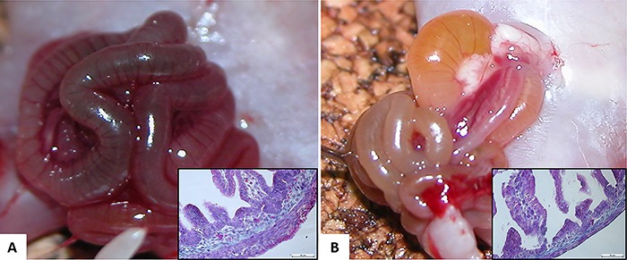
Figure 1. A, Macroscopic view of the intestinal evisceration of the gastroschisis (G) group (time of harvest). Inset: histological representation of ileum specimen stained with H&E of the G group. B, Macroscopic view of the intestinal evisceration of the gastroschisis+cannabidiol (GCBD) group (time of harvest). Inset: histological representation of the ileum specimen stained with H&E of the GCBD group. Note the different aspect and thickness of the bowel loop macroscopically and microscopically between the 2 groups. Masson's trichrome staining from ileum segment. Magnification: 100×. Scale bar: 10 μm.