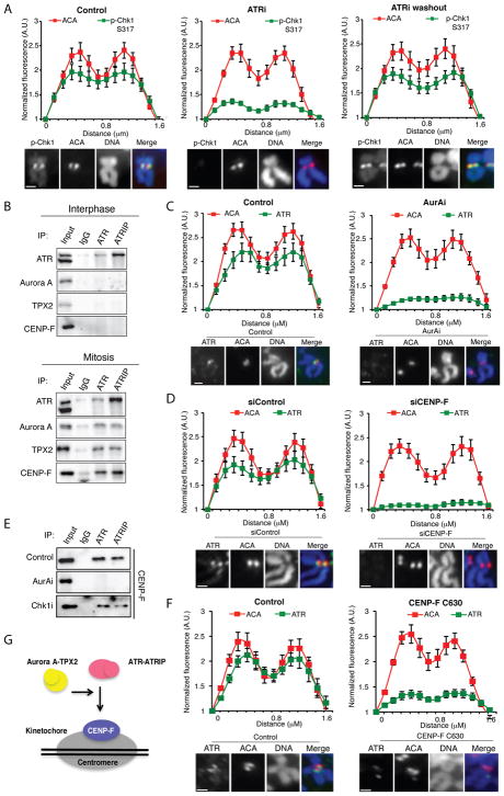
Figure 3. ATR localizes to centromeres in an Aurora A and CENP-F-dependent manner.
(A) Line scan analysis (top) and representative images (bottom) of centromeric p-Chk1 and ACA in chromosome spreads of RPE1 cells. Mitotic cells were isolated by shake off after 4h of nocodazole treatment. Cells were then mock treated (control) or treated with 2 μM VE-821 (ATRi) for 1 h in the presence of nocodazole. The ATRi washout sample was released from VE-821 for 1 h in the presence of nocodazole. (B) Immunoprecipitates of endogenous ATR and ATRIP from interphase (top) or mitotic (bottom) RPE1 cell extracts. Input = 5%. (C) Line scan analysis (top) and representative images (bottom) of centromeric ATR in chromosome spreads of RPE1 cells. Cells were arrested with nocodazole for 4h and then mock or AurAi treated for 1 h. (D) Line scan analysis of centromeric ATR and ACA in chromosome spreads of RPE1 cells. Cells were treated with control or CENP-F siRNA and arrested with nocodazole for 5 h. (E) CENP-F in the immunoprecipitates of endogenous ATR and ATRIP from mitotic RPE1 cell extracts. Cells were treated with AurAi and Chk1i as indicated. Input = 5%. (F) Line scan analysis (top) and representative images (bottom) of centromeric ATR and ACA in U2OS cells uninfected or infected with CENP-F C630 expressing retrovirus. Scale bar in all panels, 2 μm. Error bars in all panels represent SEM. *P ≤ 0.01, two-tailed t-test. (G) A model in which ATR is recruited to the vicinity of centromeres through an interaction with CENP-F. This interaction is dependent on Aurora A activity and occurs specifically in mitosis.

