Figure 3.
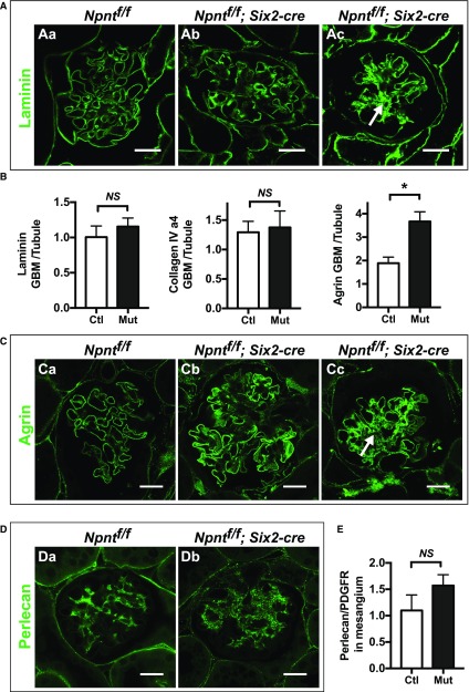
Absence of Npnt increases mesangial matrix deposition, especially in severely affected glomeruli. (A) Immunofluorescence with laminin in 4-month-old control (Ctl; Npntf/f, Aa) and mutant (Mut; Npntf/f; Six2-cre, Ab and Ac) mice. A severely affected glomerulus is shown in (Ac), which has increased laminin in the mesangial area (arrow). (B) Quantification of laminin, collagen IV (α4), and agrin in the GBM. Peak fluorescence intensity was obtained along lines drawn perpendicularly through the GBM at capillary loops in the rim of the glomerular tuft. These values were normalized to average fluorescence intensity from similar lines through the basal lamina of adjacent control tubules in the same image. Agrin is increased in the GBM of mutant mice (*P=0.003). n=3 mice of each genotype. Analysis with the unpaired t test. (C) Immunofluorescence of agrin, quantified in (3B). (Cc) A severely affected glomerulus has increased agrin in the mesangial area (arrow). (D) Immunofluorescence of perlecan in (Da) control and (Db) mutant mice. (E) Quantification of perlecan in the mesangial matrix. To attempt to normalize for differences in mesangial area, perlecan signal was normalized to PDGFRβ signal within the glomerular tuft using ImageJ (P=0.09). n=3 mice of each genotype. Analysis with the unpaired t test. Scale: 20 μm in (A, C, and D).

