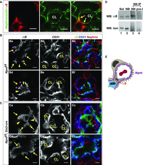
Figure 5.
Npnt localizes with α8 integrin and is required for the formation of unique GBM-mesangial adhesions. (A) Immunofluorescent imaging of α8 and Npnt in an adult mouse glomerulus shows that α8 in mesangial cells and Npnt at the GBM colocalize (arrows) at the lateral aspects of mesangial, tongue-like protrusions contacting the capillary loops (CL). (B) High-magnification images of control (Npntf/f) glomeruli show the lateral enrichment of α8 (Ba and Bd, asterisks) at mesangial protrusions (arrows), which appear to tether the base of capillary loops (CL). Other portions of the mesangial cell (Ba and Bd, arrowheads) have lower α8. CD31 and nephrin indicate the capillary loop and podocytes. (C) High-magnification images of the same structure in mutant (Npntf/f; Six2-cre) glomeruli show little focal α8 enrichment and the absence of mesangial, tongue-like protrusions (Ca and Cd, arrows) at capillary loops (CL). The α8 integrin is more diffuse (arrowheads). (D) Immunoprecipitation (IP) of anti-nephronectin from neonatal kidney lysates. Lanes: (1) Soluble lysate (Sol), the input before IP (2.5%); (2) nonbound lysate (NB), the input after IP (2.5%); (3) eluate from IP with anti-nephronectin antibody (NN); and (4) eluate from IP with preimmune antibody (pre-I). Western blots of α8 integrin and laminin are shown. Nephronectin coimmunoprecipitates α8 integrin. (E) A schematic showing the α8-enriched lateral protrusions of mesangial cells that contact nephronectin at the GBM. These appear to tether the opposing capillary loop. Scale: 4 μm in (A–C).

