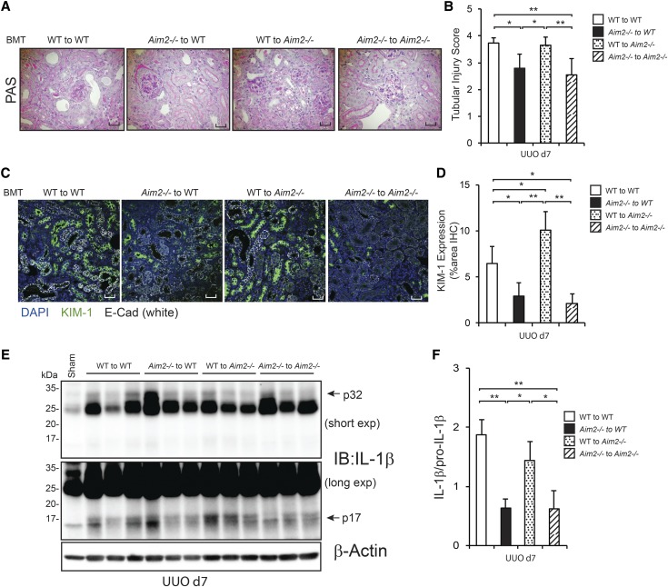
Figure 6.
Aim2 in bone marrow–derived cells contributes to tubular injury and inflammation in UUO. BMTWT to WT, BMTAim2−/− to WT, BMTWT to Aim2−/−, and BMTAim2−/− to Aim2−/− bone marrow chimera mice were generated and underwent UUO. The kidneys were harvested after 7 days (n=7, 7, 6, 5). (A) Representative photographs of periodic acid–Schiff (PAS) staining are shown. (B) Quantitative analysis of tubular injury score. (C) Immunofluorescence probing for KIM-1 (green) and E-cadherin (white) to identify tubular injury. Nuclei were contained with DAPI (blue). Representative photographs are shown. (D) Quantitative analysis of KIM-1 immunofluorescence. (E) Renal expression of pro–IL-1β and IL-1β (p17) was assessed by immunoblotting. β-actin was used as the loading control. (F) Quantitative analysis of IL-1β (p17)/Pro–IL-1β (n=3 for each). Scale bars=50 µm. Data are expressed as mean±SD, and analyzed using ANOVA with Tukey’s post hoc test. *P<0.05, **P<0.01. DAPI,4′,6-diamidino-2-phenylindole; E-Cad, E-cadherin; exp, exposure; IB, immunoblotting; IHC, immunohistochemistry.

