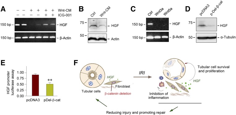
Figure 8.
Wnt/β-catenin suppresses HGF expression in vitro. (A) RT-PCR analyses show that Wnt-enriched conditioned medium suppressed HGF gene expression in cultured NRK-49F fibroblasts, whereas blockade of Wnt/β-catenin signaling by ICG-001 restored, at least partially, HGF expression in vitro. (B) Western blot analyses reveal that Wnt-enriched conditioned medium inhibited HGF protein expression in NRK-49F cells in vitro. (C) RT-PCR analyses show that recombinant Wnt3a and Wnt5a repressed HGF gene expression in cultured NRK-49F fibroblasts in vitro. (D) Western blot analyses demonstrate that constitutive activation of β-catenin also inhibited HGF expression in human cervical cancer cells (C-33A). C-33A cells were transfected with N-terminally truncated, stabilized β-catenin (pDel-β-cat) or empty vector (pcDNA3), respectively. (E) Expression of stabilized β-catenin suppressed HGF gene transcription in luciferase assay. NRK-49F cells were cotransfected with the pGL3-HGF promoter reporter plasmid (HGF promoter region, −1037 to +56) or pGL3-Basic vector, and N-terminally truncated, stabilized β-catenin expression vector (pDel-β-cat). Luciferase activity was assessed and reported as fold induction over the controls. **P<0.01 versus pcDNA3 control (n=3). (F) Schematic diagram depicts the potential mechanisms accounting for fibroblast-specific β-catenin ablation protecting against tubular injury after AKI by promoting HGF signaling. Ctrl, control.

