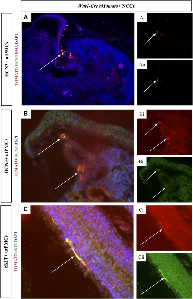
Figure 1.
HCN3+ and cKIT+ utPMCs colocalize with Wnt1-Cre;tdTomato labelled NCCs. (A and B) Lower and higher power images are shown, respectively. (A and B) TOMATO (red color, Ai and Bi) colocalizes with HCN3 (green color, Aii and Bii) generating orange/yellow color in the merged image in PKJ cells adjacent to the renal artery (A and B, arrows). (C) TOMATO (red color, Ci) colocalizes with cKIT (green color, Cii) generating orange/yellow color in the merged image in thin, elongated cells adjacent to the ureteric wall (C, arrows). cKIT, KIT Proto-Oncogene Receptor Tyrosine Kinase; DAPI, 4′,6-Diamidino-2-Phenylindole; HCN3, Hyperpolarization Activated Cyclic Nucleotide Gated Potassium Channel 3; NCC, neural crest cell; SMA, smooth muscle actin; utPMCs, urinary tract pacemaker cells.

