Figure 5.
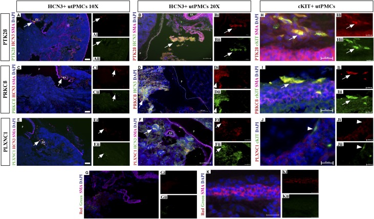
Novel utPMC markers are enriched in HCN3+ and cKIT+ utPMCs at the protein level in situ. Immunofluorescence analysis revealed colocalization between PTK2β (green, A–Aii; red B–Bii), PRKCβ (green, C–Cii; red, D–Dii), PLXNC1 (green, E–Eii; red, F–Fii), and HCN3 (green, A, C, and E; red, B, D, and F). Colocalization was observed in cells adjacent to the renal artery (RA) outside the pelvis (P) consistent with the location of utPMCs. (G–Gii) Control IgG sections revealed weak/no signal in the red and green channels. Analysis revealed colocalization between cKIT (green, H–Jii), and PTK2β (red, H–Hii) or PRKCβ (red, I–Iii), but not with PLXNC1 (red, J–Jii). cKIT staining was observed in elongated cells in the adventitial layer adjacent to the ureteric muscular layer consistent with the localization of cKIT+ utPMCs. (K–Kii) Control IgG sections revealed weak/no signal in the adventitial layer in red and green channels. Scale bars represent 200 µm (A, C, and E), 100 µm (B, D, F, and G), or 20 µm (H–K). cKIT, KIT Proto-Oncogene Receptor Tyrosine Kinase; DAPI, 4′,6-Diamidino-2-Phenylindole; HCN3, Hyperpolarization Activated Cyclic Nucleotide Gated Potassium Channel 3; PLXNC1, Plexin C1; PRKCβ, Protein Kinase C Beta; PTK2β, Protein Tyrosine Kinase 2 Beta; SMA, smooth muscle actin; utPMCs, urinary tract pacemaker cells.

