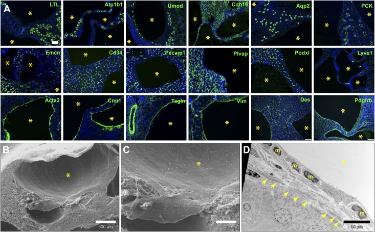
Figure 3.
Renal cysts arising from loss of Angpt1/2-Tie signaling uniformly express myofibroblast markers. (A) Representative immunofluorescence analysis of renal cysts (yellow asterisks) found adult A1/A2ΔE16.5mutants showing lack of expression of epithelial tubule–specific (L. tetraglobulus lectin [LTL], Na+/K+-ATPase [Atp1b1], uromodulin [Umod], Ksp-cadherin [Cdh16], aquaporin-2 [Aqp2], and pancytokeratin [PCK]) and endothelial (Emcn, Cd34, Pecam1, Plvap, Podxl, and Lyve1) markers but strong expression of myofibroblast markers (Acta2, Cnn1, Tagln, Vim, desmin [Des], and PDGFRβ receptor [Pdgfrb]). (B and C) Scanning electron and (D) transmission electron micrographs of renal cysts (yellow asterisks) in an adult Tie2ΔE16.5 kidney showing the squamous mesenchymal morphology of cells lining cysts (nuclei labeled m). Fibrillar deposits are also visible underneath the cyst linings (arrows). Scale bars, 100 μm in A; 500μm in B; 200 μm in C; 10 μm in D.

