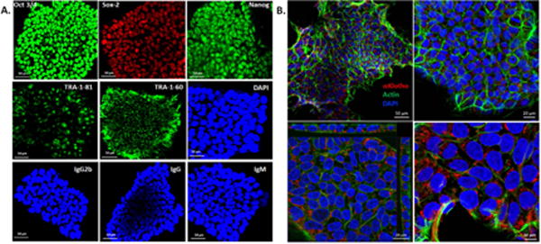
Figure 1.

A. Immunophenotypic profile of iPSC colonies. Following viral transfection of fibroblasts and culture × 2 to 3 weeks, iPSC colonies stained positive for pluripotency markers OCT3/4, Sox-2, NANOG, TRA-1-60 and TRA-1-81. DAPI stains the nuclei. The colonies were also stained for isotype control antibodies IgG2b (Oct3/4), IgG (Sox-2 and Nanog), and IgM (TRA-1-81 and TRA-1-60). Bar=50μm. B. iPSC colonies imaged at different magnifications (upper 2 panels) show abundant expression of αKlotho (red), counterstained for filamentous actin (Alexa Fluor 488 phalloidin, green) and nuclei (DAPI, blue) Bar = 50 μm (left) and 20 μm (right). Z-section (left lower panel) shows the cuboidal nature of cells and further magnified (right lower panel) to show αKlotho in the cytoplasm. Bar = 20 μm (left) and 10 μm (right).
