
Figure 6.
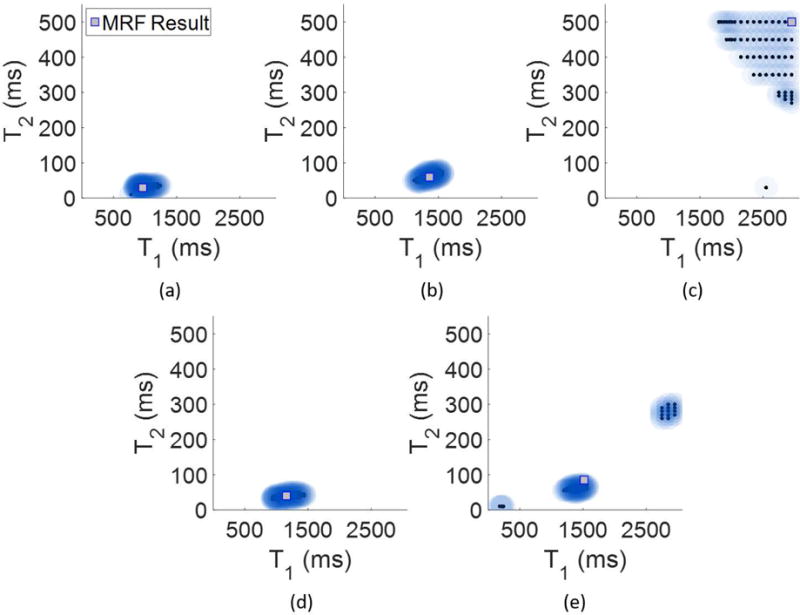
Voxel-wise algorithm results from the patient, diagnosed with a GBM. Panel (a) shows the result from a pure white matter voxel, (b) from a pure gray matter voxel and (c) from a pure CSF voxel. Panels (d) and (e) show Bayesian MRF results from two voxels which potentially exhibit partial volume. In (d) is a mix of white matter and gray matter, and in (e) is a mix of gray matter and CSF.