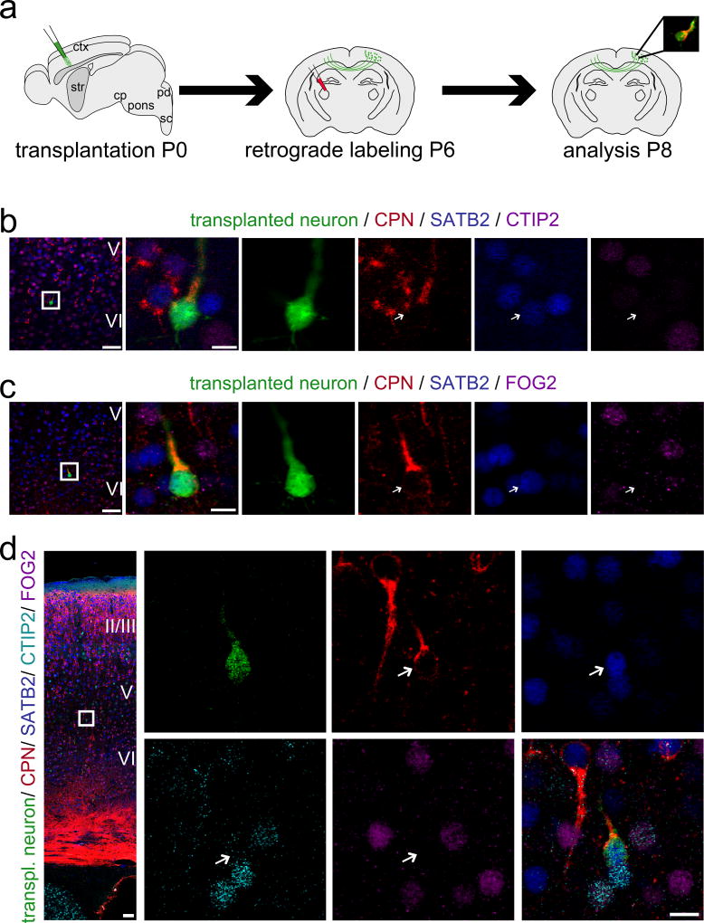
Figure 5. Newly incorporated neurons of combinatorial callosal molecular identity establish anatomic inter-hemispheric output connectivity with high fidelity.
(a) Micro-transplantation of eGFP+ single cell suspension into P0/P1 recipient sensorimotor cortex. Stereotaxic injection of retrograde axonal tracer (CTB555) into the contralateral hemisphere at P6. Analysis at P8. (b, c) 4-channel confocal analysis. (d) Five-channel/spectral unmixing confocal analysis. eGFP+ and trans-callosally projecting transplanted neurons express the callosal marker SATB2 and are negative for the subcerebral marker CTIP2 (b, d) and for the corticothalamic marker FOG2 (c, d). Arrows indicate transplanted neurons. The positions of the boxes in low magnification panels (b – d) correspond to the cortical area examined in high magnification panels. (b): eGFP (green), CTB555 (red), SATB2 (blue), CTIP2 (purple); (c): eGFP (green), CTB555 (red), SATB2 (blue), FOG2 (purple); (d): eGFP (green), CTB555 (red), SATB2 (blue), CTIP2 (cyan), FOG2 (purple). Scale bars: b – d (low magnification), 50 µm; b – d (high magnification), 10 µm. Molecular identity data are presented for three transplanted and retrogradely labeled CPN; primary data from six additional neurons are presented in Fig. S7. Taken together, all analyzed transplanted and trans-callosally projecting neurons possess CPN molecular identity, and no transplanted neurons of SCPN or CThPN molecular identity were retrogradely labeled from the contralateral hemisphere.

