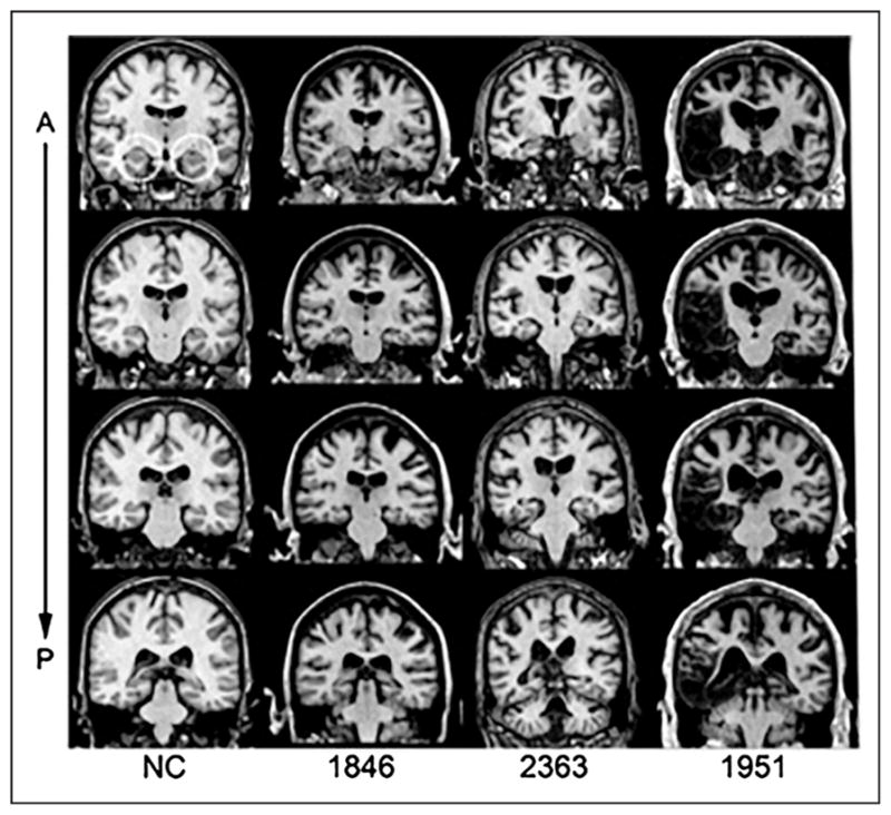
Figure 1.

Magnetic resonance scans of hippocampal patients. Images are coronal slices through four points along the hippocampus from T1-weighed scans. Volume changes can be noted in the hippocampal region for Patients 1846 and 2363, and significant bilateral MTL damage including the hippocampus can be noted in Patient 1951. A = anterior; P = posterior; NC = healthy comparison brain.
