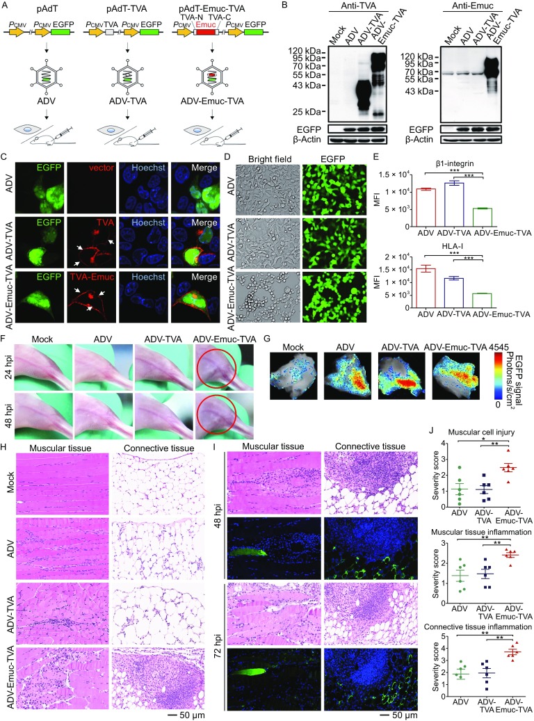
Figure 1.

Adenoviral vector-mediated gene delivery and the potential pathogenicity analyses of EBOV mucin-like glycoprotein (Emuc) in vitro and in vivo. (A) Technological process of the present research. Firstly, the cassettes encoding the small cell membrane protein TVA or the fused Emuc-TVA were respectively constructed into the donor plasmid pAdT which contains an EGFP expression cassette as the reporter. Adenoviral vectors were then generated by homologous recombinant in bacteria and packaging in HEK293 cells. After identification and purification, the recombinant viral vectors were utilized in the following gene transfer to cultured cells in vitro and mouse muscles in vivo. (B) Identification of the expression of the cloned genes. HEK293 cells were mock infected or infected by the recombinant adenoviruses ADV-Emuc-TVA, ADV-TVA, or ADV. At 24 hpi, cell lysates were analyzed by Western-blot with the anti-TVA or anti-Emuc antibodies, respectively. EGFP expression was detected for monitoring the efficient and comparable transduction; β-actin was included for sample loading control. The detection was repeated for at least three times with similar results. The multiple bands likely represent the various glycosylation forms of the proteins. (C) Protein subcellular localization. Vero cells infected with the indicated recombinant adenoviruses were fixed and permeabilized at 24 hpi for IFA using the anti-TVA antibody and the localization of target proteins was then visualized under confocal microscope. White arrows indicate the substantial cell membrane localization. See also Fig. S1. (D) Emuc induces rounding and detachment of cultured adherent cells. Adherent HEK293A cells were infected by the indicated viral vectors. At 24 hpi, viral infection (as indicated by EGFP expression) and cellular morphological changes were monitored by a Nikon microscope. A representative result from at least three-time independent experiments was shown. (E) HEK293A cells infected with the indicated viruses were fixed at 36 hpi for surface immunostaining of the cell membrance molecules, β1-integrin or HLA-I, using the corresponding antibodies respectively. Fluorescence signals of the surface molecules were detected by flow cytometry and mean fluorescence intensity (MFI) was analyzed. Graphs show mean ± SD, n = 3. ***, P < 0.001. (F) BALB/c mice were divided into 4 groups randomly and mock infected or infected with the indicated viruses by intramuscular injection (i.m). Clinical feature then was monitored daily. Circles indicate redness and inextensibility of the hind limb muscles at 24 and 48 hpi, respectively. (G) The mice infected as indicated were euthanized at 48 or 72 hpi, respectively. EGFP signals of the dissected skeletal muscles were detected by an animal imaging system. Representative samples at 72 hpi were shown. (H) Paraffin sections of the indicated skeletal muscles (72 hpi) were stained with H&E for histopathological analyses. See also Figs. S3 and S4. (I) Continuous slices of the paraffin-embedded sample infected by ADV-Emuc-TVA were subjected to H&E staining and IFA for the analyses of histopathology and Emuc-TVA expression, respectively. (J) Quantification of histopathological changes. Unduplicated images of muscular tissues and connective tissues for each sample (72 hpi) as indicated were chosen, respectively, and the degrees of pathological changes in comparison to mock-infected samples were scored by the standard: 0 = normal; 1 = minimal change; 2 = mild change; 3 = moderate change; 4 = marked change; 5 = severe change. Graphs show mean± SD, n = 6. **, P <0.01; *, P < 0.05. See also Fig. S5
