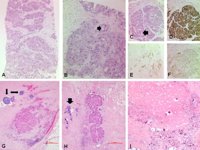
Fig. 4.
A–F. Liver biopsy of fibrolamellar hepatocellular carcinoma (FL-HCC): A Solid nests of large tumour cells with abundant eosinophilic cytoplasm and hyperchromatic or vesicular nuclei and prominent nucleoli are seen within a lamellar fibrous stroma (haematoxylin and eosin—H&E, A ×10, B ×20, C ×20). Intra-cytoplasmic pale bodies (black arrow) are abundant. The FL-HCC cells are typically positive for HepPar1 (D ×20), keratin 7 (E ×20) and CD68 (F ×20). G, I Resection specimen following TACE and SIRT treatment: G, H isolated, less than 2-mm sized foci of residual viable FL-HCC (H&E ×4) surrounded by TACE spheres (black arrows). I Coagulative tumour necrosis with cholesterol clefts, pigmented macrophages and TACE spheres (H&E ×4)

