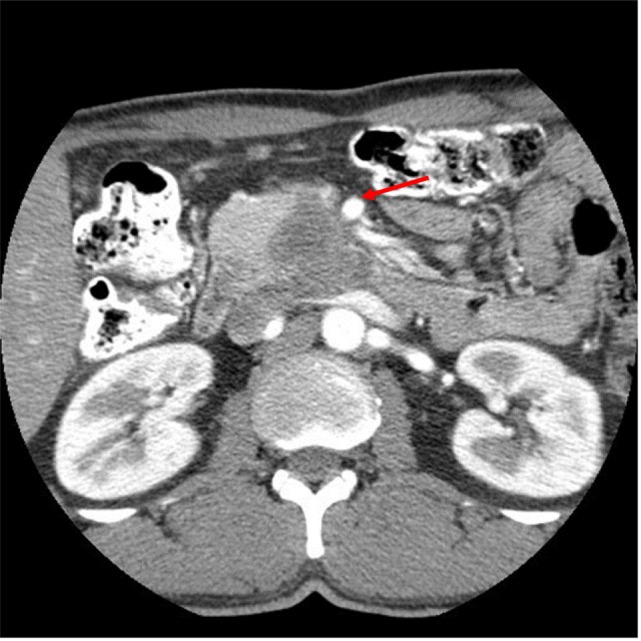
Figure 1.

CT abdomen at initial work-up that shows a hypo-attenuated mass in the region of the uncinate process contacting 50% of the superior mesenteric vein, and approximately 25% of the superior mesenteric artery (see arrow) as well as contacting the left renal vein.
