Figure 1.
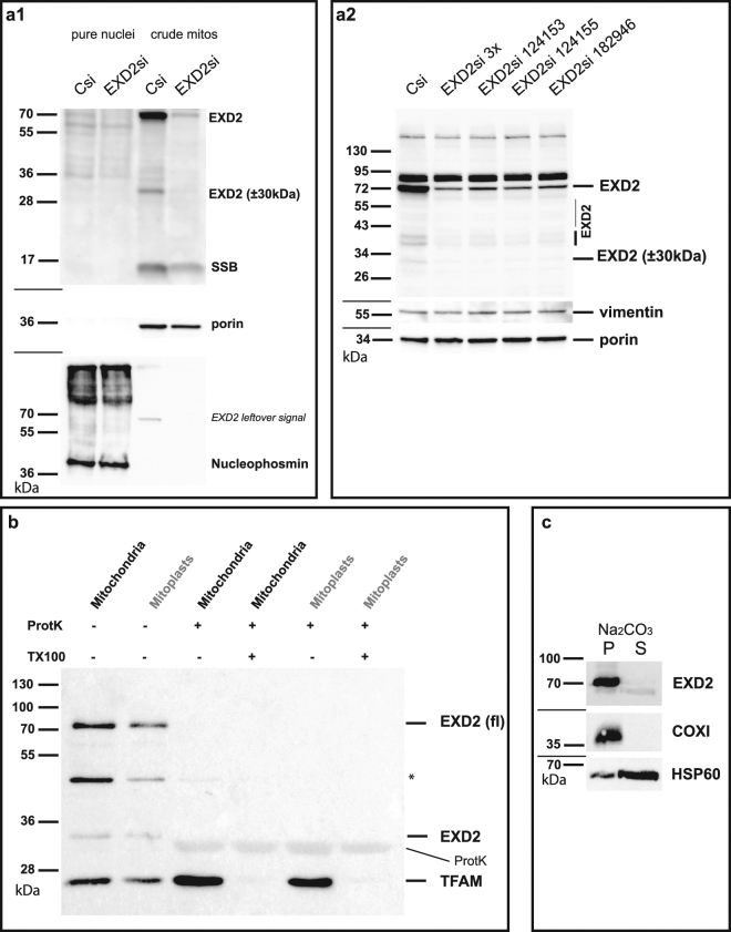
EXD2 is a mitochondrial outer-membrane associated protein. U2OS cell crude mitochondria isolated by differential centrifugation and pure nuclei isolated on iodixanol gradients were tested for EXD2 abundance in control siRNA and EXD2 siRNA treated cells (panel a1). Results show that the vast majority of EXD2, similar to mitochondrial marker proteins mtSSB and porin are found in the mitochondrial fraction and not in the nuclear fraction, in which the nuclear marker nucleophosmin is identified. SiRNA treatment confirms the identity of the full length (fl) ~70 kDa EXD2 protein and several lower abundant species of 30 kDa and higher molecular weight. Knockdown with either the combined pool of three commercial siRNAs or each individual siRNA show similar knockdown efficiencies in total cell lysates of U2OS cells on Western blots (panel a2). At the same time, several lower molecular weight EXD2 species are also identified and all appear to be equally sensitive to each individual siRNA suggesting they might be breakdown or processed EXD2 forms. Protease protection demonstrates mitochondrial EXD2 is mostly found in the mitochondrial outer-membrane (panel b). Mitochondria and digitonin-derived mitoplasts from HEK293 cells were treated either with Proteinase K (ProtK) alone or with ProtK and Triton-X100 (TX100) to lyse the inner and outer membrane. Results show that whereas TFAM, an mtDNA associated protein, is protected from ProtK in the absence of TX100, EXD2 is not, suggesting and outer-membrane localization. *Indicates a remnant signal from the probing with an antibody against a different mitochondrial candidate protein not relevant for this paper. A Na2CO3 extract of crude mitochondria from HEK293 cells (panel c) shows that similar to cytochrome c oxidase subunit I (an integral membrane protein), full length EXD2 is found predominantly in the pellet (membrane) fraction, whereas the majority of HSP60 is found in the supernatant (non-membrane) fraction. For each panel (except panel b) cropped images show the results of incubations with subsequent antibodies on the same blots, indicated by dividing lines (see Supplementary info for full blot images).

