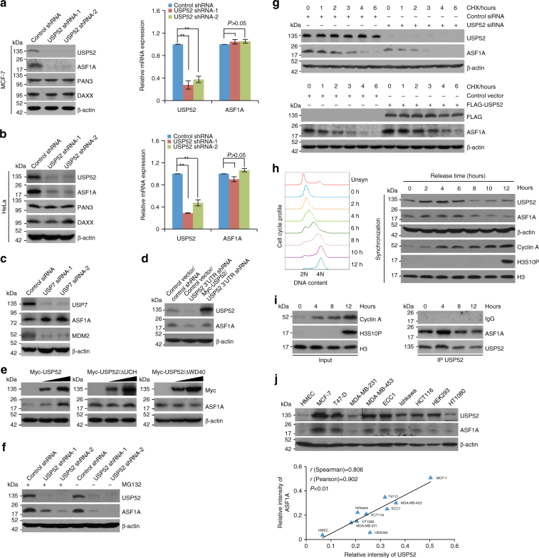
Fig. 4.
USP52 promotes ASF1A stabilization. a MCF-7 cells stably expressing different sets of USP52 shRNAs were collected for western blotting and qRT-PCR analysis. Each bar represents the mean ± S.D. for biological triplicate experiments. **P < 0.01, one-way analysis of variance (ANOVA). b Experiments analogous to a were performed in HeLa cells. Each bar represents the mean ± S.D. for biological triplicate experiments. **P < 0.01, one-way ANOVA. c MCF-7 cells were transfected with control siRNA or USP7 siRNA. Cellular extracts were prepared and analyzed by western blotting. d MCF-7 cells stably expressing shRNA targeting 3′UTR of USP52 were transfected with control vector or Myc-USP52 and cellular lysates were collected for western blotting analysis with antibodies against the indicated proteins. e MCF-7 cells were transfected with different amounts of wild-type USP52, USP52 lacking UCH domain (USP52/∆UCH), or USP52 lacking WD40 repeat domain (USP52/∆WD40), and cellular lysates were collected for western blotting analysis with antibodies against the indicated proteins. f MCF-7 cells stably expressing different sets of USP52 shRNAs were treated with proteasome inhibitor MG132 (10 μM) or DMSO. Cellular extracts were prepared and analyzed by western blotting. g MCF-7 cells transfected with control siRNA or USP52 siRNA were treated with cycloheximide (CHX) and harvested at the indicated time followed by western blotting analysis (upper panel). MCF-7 cells stably expressing control vector or FLAG-USP52 were treated with CHX and harvested at the indicated time followed by western blotting analysis (lower panel). h HeLa cells synchronized by double-thymidine block were released and cellular extracts were collected for western blotting analysis with antibodies against the indicated proteins. Representative cell cycle profiles are shown. i HeLa cells synchronized by double-thymidine block were released and cellular extracts were collected for co-immunoprecipitation analysis of the association of ASF1A with USP52. j Western blotting analysis of the expression of ASF1A and USP52 in multiple cell lines. The intensity of each band was quantified by densitometry with Image J software with β-actin as a normalizer. The correlation coefficient and P-values are shown

