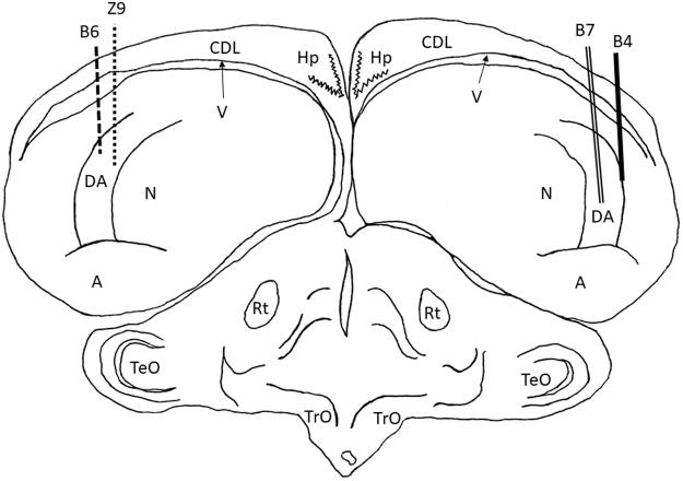
Figure 2.
Electrode track reconstruction. Electrode track position reconstructions for the two right NCL birds (B4 and B7) and two left NCL birds (B6 and Z9). All recordings were within the full dorsal-ventral extent of NCL. The following are the brain regions as defined by Reiner et al.26: A, archopallium; DA, tractus dorso-arcopallialis; CDL, area corticoidea dorsolateralis; Hp, hippocampus; N, nidopallium; Rt, nucleus rotundus; TeO, tectum opticum; TrO, tractus opticus; V, ventricle.

