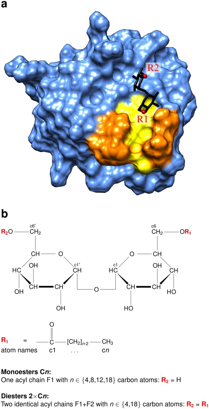
Figure 1.

Structure of Mincle and the glycolipid ligands. (a) Structure of the Mincle carbohydrate recognition domain (PDB code: 4ZRV; ref.7). The protein domain is shown in surface presentation and the hydrophobic groove representing a putative ligand binding site is highlighted in yellow. Residues L172, V173, F197, and F198 that line the hydrophobic groove are marked in orange. The trehalose moiety is shown as stick presentation and the attachment sites of the first and second acyl chain are labelled R1 and R2, respectively. (b) Structure of trehalose acyl esters. Trehalose is a disaccharide consisting of two glucose moieties. Atoms are referred to by lower case characters in the present study. Monoesters are esterified at the c6 atom, diesters both at the c6 atom of the first and the c6′ atom of the second glucose moiety.
