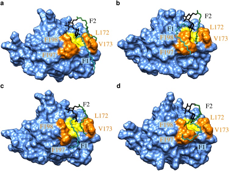
Figure 7.
Representative binding modes of the 2 × C18 diester to Mincle. Mincle is shown in blue surface presentation with the hydrophobic groove in yellow. Hydrophobic residues lining the groove are shown in orange and are labelled. The glycolipid is shown in stick presentation with the trehalose, F1 and F2 chains are colored in black, cyan, and green, respectively. See text for a detailed description of the binding modes (a–d) and the Supplementary movie for the dynamic nature of the interaction.

