Figure 4.
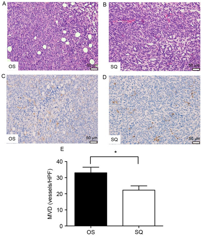
H&E staining and immunohistochemical staining analysis of primary tumors (magnification, ×200) with (A) OS and (B) SQ implantation. Representative CD31 staining of primary tumors with (C) OS implantation and (D) SQ implantation. Brownish staining indicated CD31+ vessels. (E) Quantification of MVD as numbers of CD31+ vessels/field (magnification, ×100; *P<0.05). H&E, hematoxylin and eosin; CD, cluster of differentiation; OS, orthotopic implantation; SQ, subcutaneous implantation; MVD, microvessel density; HPF, high-power field.

