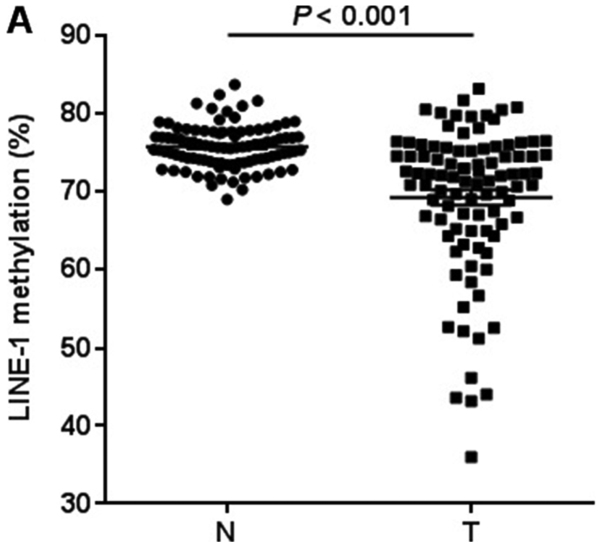
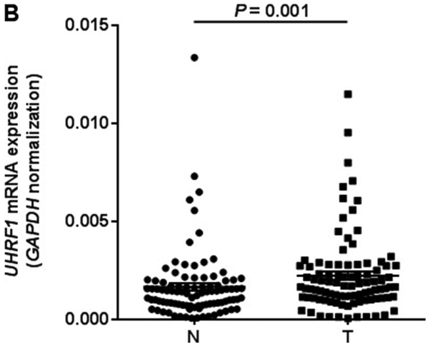
Figure 2.
LINE-1 methylation and UHRF1 expression comparision between 95 gastric cancer tissues and matched adjacent normal tissues. (A) LINE-1 methylation levels. The cancer tissues showed significantly lower levels of methylation than matched normal tissues (P<0.001 by Wilcoxon signed-rank test). (B) UHRF1 expression levels. UHRF1 mRNA expression levels were significantly higher in cancer tissues than matched normal tissues (P=0.001 by Wilcoxon signed-rank test). The mRNA levels were normalized to those of GAPDH. Horizontal lines represent the mean ± standard error of the mean as determined by triplicate assays. N, adjacent normal tissues; T, tumor tissues; LINE-1, long interspersed element-1; UHRF1, ubiquitin-like with PHD and ring-finger protein 1.


