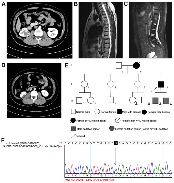
Figure 3.
Examination of case 3. (A) CT scan indicating multiple nodules with low-density shadows in left and right kidneys. The larger nodule in the right kidney had a diameter of ~34 mm. Contrast-enhanced scanning showing uneven enhancement in a progressive manner. Bilateral renal and pancreatic multiple cysts were observable. (B) Contrast-enhanced MRI scan showing minor enhancement, which was indicative of a hemangioma. A round mixed signal (diameter, ~12 mm) was observable in the sixth thoracic vertebra. (C) MRI scan showing a round long T2 signal in the horizontal sacral area of S2, which indicated the presence of a sacral cyst. (D) Contrast-enhanced CT scanning revealed no marked enhancement. Multiple cystic lesions were observed in the left and right kidneys. The lower part of the right kidney was altered following surgery. (E) Pedigree chart of case 3. (F) Genetic test results in case 3 confirmed by Sanger sequencing. CT, computed tomography; MRI, magnetic resonance imaging; VHL, von Hippel-Lindau.

