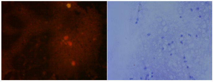
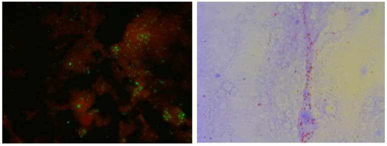
Figure 1.
Brain impression from a non-rabid dog (top two panels) or a dog suspected of rabies (bottom two panels), subjected to direct fluorescent antibody (DFA) test using anti-rabies virus nucleocapsid protein IgG-FITC conjugate (left two panels) or to direct rapid immunochemistry test with (dRIT) using biotinylated mouse anti-rabies monoclonal antibodies and streptavidin-peroxidase, with hematoxylin couterstain (right two panels). Scale: 200×.


