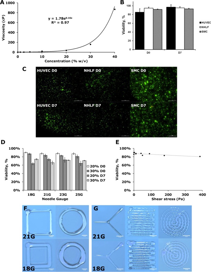
Figure 6.
Cytotoxicity and printability of elastic PEG–PCL(24k)-DA hydrogel using an extrusion bioprinter. (A) Viscosity curve of elastic PEG–PCL(24k)-DA precursor solution. (B, C) Cell viability of different cell types in printed 10% elastic PEG–PCL(24k)-DA hydrogel. Live/dead assay was performed immediately after gel polymerization and after 7 days in culture (scale bars represent 500 μm). (D) Effect of different needle sizes and precursor solution concentrations on viability of neonatal human lung fibroblasts. (E) Effect of shear stress on cell viability evaluated immediately after printing. (F, G) Sample shapes printed using a printer with different needle sizes (scale bars in (F) represent 2 mm and scale bars in (G) represent 5 mm).

