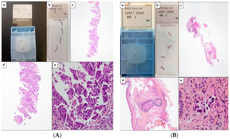
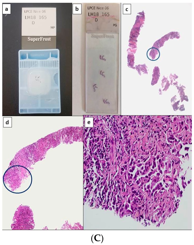
Figure 1.
Potential scenarios for detection of theranostic biomarkers for lung adenocarcinomas using thoracic biopsies. Depending on the quantity of tumor cells detected on hematoxylin and eosin stained sections, immunohistochemistry (IHC) and/or molecular biology approaches can be used (with or without panels). (A) Large biopsy with a high percentage of tumor cells (more than 50%). Paraffin block with one biopsy (a) and corresponding 4 tissue sections stained with hematoxylin eosin (b). Different magnifications of the same tissue section showing high number of adenocarcinoma cells (c–e). In this latter case, the number of tumor cells should allow PD-L1 IHC to be performed first and then followed by NGS. Alternatively a workflow including successive PD-L1, ALK, ROS1, and BRAF IHC, and then NGS can be also adopted. (B) Small biopsies with a few tumor cells. Paraffin block with small biopsies (a) and corresponding 4 tissue sections stained with hematoxylin eosin (b). Different magnifications of the same tissue section showing in only one area (circle) a low number of tumor cells (no more than 15 tumor cells) (c–e). IHC targeting PD-L1, then IHC for EGFR, ALK, ROS1, and BRAF should be probably done. (C) Large biopsies with a low percentage of tumor cells (less than 5%). Paraffin block with at least 5 large biopsies (a) and corresponding 4 tissue sections stained with hematoxylin eosin (b). Different magnifications of the same tissue section showing in only one area (circle) some tumor cells (c–e). IHC for PD-L1 then successively for ALK, ROS1 and BRAF can be done and followed by targeted molecular biology for detection of EGFR mutations on a single tissue section or alternatively IHC for EGFR, according to the sensitivity of the MB test [1Ac, 1Bc, 1Cc, original magnification ×25; 1Ad, 1Bd, 1Cd, original magnification ×100; 1Ae, 1Be, 1Ce, original magnification ×400].


