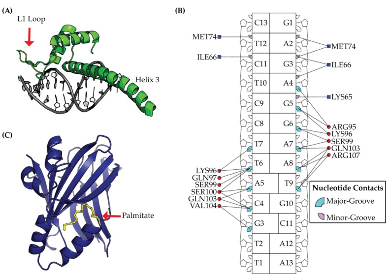
Figure 3.
TEAD is a protein with multiple domains, which is composed of a DNA Binding Domain (DBD) and a YAP Binding Domain (YBD). (A) The DBD structure is illustrated from protein data bank ID (PDB) 5GZB and is composed of a homeodomain fold with three alpha helices (shown in green) bound to DNA (colored in grey); (B) Structural analysis of TEAD DBD using the interaction map from DNAproDB [34] illustrates the DNA major- and minor-groove protein residue contacts in cyan and pink, respectively. Nucleic acid contacts with DBD L1 loop are indicated by a blue square and contacts with DBD helix 3 are shown as red circles; (C) The YBD is post-translationally modified by palmitate (colored yellow) that extends towards the interior of YBD (colored in blue).

