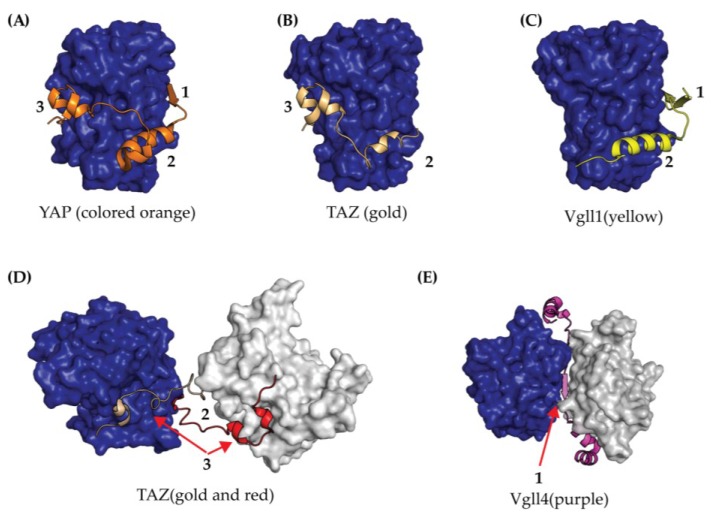
Figure 4.
Crystal Structures of TEAD YBD (colored blue) bound to co-activator peptides from (A) YAP (orange); (B) TAZ binding mode one (gold); (C) Vgll1 (yellow); (D) TAZ binding mode two (gold and red); and (E) Vgll4 (purple). In both (D) and (E), the YBD is shown in blue and grey surface renderings since co-activators in the crystal structure induced dimerization. The TEAD binding interfaces for each co-crystal structure are labeled as 1, 2, and 3.

