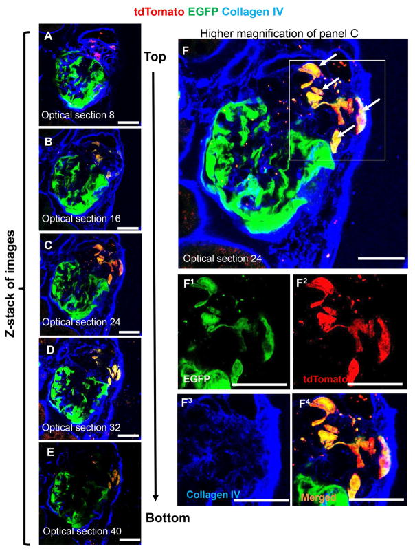
Figure 3. A subset of tdTomato+ CoRL migrate onto the glomerular tuft and begin to co-express EGFP.
Immunohistochemistry for Collagen IV is used to easily identify glomeruli.
(A–E) Fifty 0.4 μm optical sections were taken by confocal microscopy through a 20μm thick section of a glomerulus from CoRL-PODO mice with FSGS. Five equally distributed representative optical sections are shown. F) Higher magnification of optical section 32 (panel D) showing tdTomato+ EGFP+ co-labeled cells on the glomerular tuft, which appears yellow in merged images (arrows). Images of the areas of interest outlined by the white boxes are shown below in superscript 1–4 for green, red, far red and merged fluorescence channels respectively. Scale bars = 25μm.

