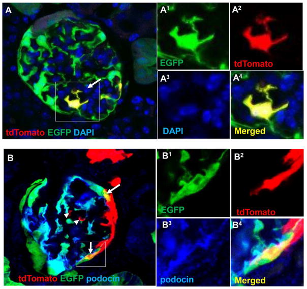
Figure 4. tdTomato+ CoRL on the glomerular tuft co-express EGFP and stain for podocyte marker Podocin.
A) Another clear example of a single tdTomato+ EGFP+ CoRL on the glomerular tuft (arrow). B) Immunohistochemistry for the podocyte protein podocin (blue) shows co-labeling with tdTomato+ EGFP+ CoRL on the glomerular tuft (arrows). Images of the areas of interest outlined by the white boxes are shown to the right in superscript 1–4 for green, red, far red and merged fluorescence channels, respectively. It should be noted that the tdTomato and EGFP reporters are not targeted to any particular cellular compartment, whereas staining for native proteins such as podocin are localized to specific structures such as the slit daphragms of foot processes in normal podocytes. 24 Furthermore, not all tdTomato+ CoRL that migrate onto the tuft (arrow heads) express podocyte markers9. Scale bars = 25μm.

